Knee Cartilage Injury Specialist
Do you have pain, swelling, or decreased range of motion in your knee? Maybe you also feel like something is moving around inside the knee joint. You may have a knee cartilage injury. Cartilage injuries specialist Dr. Prem N. Ramkumar provides non-surgical and surgical treatments for patients with cartilage injuries. His office is located in Long Beach and serves Los Angeles, Orange County, and surrounding Southern California areas. Contact Dr. Ramkumar’s team for an appointment today!

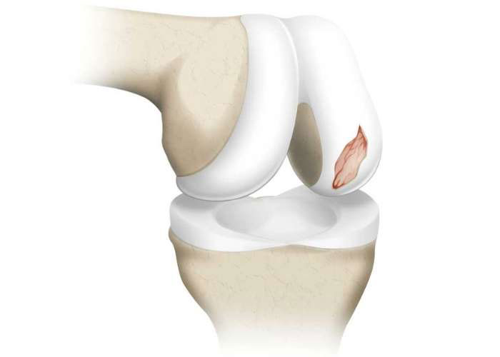
What are articular cartilage injuries of the knee?
Cartilage is found throughout your body, acting as a cushion between the bones of the joints. When the cartilage is healthy, our bones move and glide freely without any pain. When the cartilage is damaged or degenerates, the knee no longer moves freely, causing pain. Specific to the knee, two types of cartilage exist:
- Articular cartilage: Covers the undersurface of the bones
- Fibrocartilage (Meniscus): Located between the bones and acts as a cushion or shock absorber.
Articular cartilage is very tough, much like the cartilage (gristle) on the ends of chicken bones. It can maintain itself through the nutrients it receives from knee joint fluid. However, articular cartilage has no blood or nerve supply. This means that it has a very poor ability to heal. This also means that it can be subtle to realize when your articular cartilage is injured. Without treatment, focal cartilage injuries may lead to degeneration and osteoarthritis (diffuse articular cartilage injury) of the joint. Dr. Prem Ramkumar, knee cartilage injuries specialist, is located in Long Beach and serves the Los Angeles, Orange County, and surrounding Southern California areas.

How do knee cartilage injuries occur?
Trauma, knee instability, and degeneration are all ways knee cartilage injuries occur. A direct blow to the knee in sports such as hockey, football, and lacrosse is a common way athletes damage the articular cartilage. Cartilage injuries can also occur simultaneously with patellar dislocations and ligamentous injuries like the anterior cruciate ligament (ACL). The articular cartilage also wears down over time due to overuse or aging.
What are the symptoms of a knee cartilage injury?
Cartilage injuries of the knee have similar symptoms, regardless if it is an articular cartilage injury or a meniscus injury. Symptoms may include:
- Pain: Often sharp at the time of injury or a slow onset for degenerative conditions
- Swelling: Especially after activity
- Decreased range of motion
- Inability to stand or walk normally.
Sometimes a fragment of the articular cartilage breaks away and starts floating freely in the synovial fluid inside the knee joint. Known as a ‘loose body’, this can affect knee movement and often feels like something is moving inside the knee joint. If this is due to a new or acute presentation, this should be evaluated urgently (within a week).
How are knee cartilage injuries diagnosed?
An MRI scan is the best way to diagnose focal cartilage images and determine the size and shape of the injury. The presence of associated bone marrow edema is helpful to appreciate as well. X-rays can also be used to rule out diffuse articular cartilage injury (osteoarthritis), which alters the treatment plan. In certain circumstances, Dr. Ramkumar may perform a knee arthroscopy using a tiny camera called an arthroscope to more accurately characterize the lesion and address any other injuries. It will be placed inside the joint, allowing Dr. Ramkumar to display images of the knee on a monitor to determine the extent of any damage to the articular cartilage.
How are knee cartilage injuries graded?
Knee cartilage injuries are graded on a scale of 1 through 4, with 4 being the most severe.
- Grade 1: Some softening and inflammation
- Grade 2: Partial loss of thickness and some surface fissures possible
- Grade 3: High-grade partial thickness cartilage loss – defect of the subchondral bone measuring 1.5cm or less
- Grade 4: Full thickness cartilage loss, subchondral bone fully exposed.
What are the treatment options for knee cartilage injuries?
Non-Surgical treatments:
Some patients with knee cartilage injuries will respond well to conservative, non-surgical treatments that may include:
- RICE (Rest, Ice, Compression, and Elevate)
- NSAIDS (non-steroidal anti-inflammatory drugs)
- Physical therapy
- Steroid injections
- Biological injections consisting of hyaluronic acid (gel), platelet-rich plasma (PRP), bone marrow blood, or adipose (fat) based stem cells
Surgical Treatments:
Focal articular cartilage lesions can be treated in a variety of different ways depending on the age of the patient, size of the lesion, preoperative MRI findings, and intraoperative appearance. Therefore, Dr. Ramkumar may recommend one or more surgical techniques based on your age, activity level, and size and type of tear. Surgical treatments may include:
- Debridement: Smoothing the damaged cartilage and remove loose bodies
- Microfracture: Small holes are made in the bone, causing it to bleed so new cartilage forms. This was a popularized and cost-effective treatment option in the past but has since been found to be a poor surgical option with unacceptably high failure rates after 2 years.
- BMAC: Biologics in conjunction with cartilage repair. Bone marrow from the patient’s pelvis provides growth factors that may result in a cartilage-like surface.
- MACI (matrix associated autologous chondrocyte implantation): Two-stage procedure that involves harvesting your own cartilage, growing it in the lab for 6-8 weeks to create a scaffold patch, then re-implanting it over the damaged cartilage surface with glue. The cells and scaffold facilitate the creation of durable cartilage repair tissue.
- Mosaicplasty/OATS: Osteochondral autograft transfer (OATS), mosaicplasty, or autologous osteochondral transfer (AOT) are the same procedure. These terms describe a minimally invasive procedure that transfers cartilage from a non-weight bearing region of the knee to the area of injury. Typically, two-four plugs can be harvested and used to treat a symptomatic lesion of the knee condyles, trochlea or patella. Plug diameters range from 6-10 mm; plug length is usually 10-15 mm. This is usually reserved for small to medium size lesions that are 2-4 cm2. Dr. Ramkumar places these plugs to fully resurface the area of cartilage damage while backfilling the harvest site with bone that heals reliably. The benefit of this procedure is that it is single stage and the defect is immediately filled. The results of autograft osteochondral transfer are very durable. If possible, this is the best surgical option in terms of surgically managing focal articular cartilage lesions.
- Osteochondral allograft: Cartilage repair surgery that uses a donor source to transplant cartilage and bone to repair a focal articular cartilage defect is referred to as OCA. In these cases, a donated condyle specimen is used to craft a graft to resurface the knee. Typically, one or two cylindrical bone-cartilage grafts are used to restore the damaged cartilage area. These donated grafts are press fit into the defect to immediately reconstruct the joint. The donated specimens are fresh, which means that the transplanted cartilage is viable and durable. Like mosaicplasty, this minimally invasive procedure is performed in a single stage and offers immediate management of the cartilage lesion. This is usually reserved for larger lesions that are larger than 4 cm2
- Particulated Juvenile Articular Cartilage: Patellar lesions are best amenable to the implantation of small pieces of juvenile articular cartilage, which may allow for durable cartilage repair tissue in a chondral defect. This product (known as DeNovo NT, Zimmer) is comprised of viable pieces of articular cartilage from young donors, but long-term data is still lacking. However, early results have been promising.
One of the most important surgical aspects of cartilage repair surgery is understanding and appreciating the patient’s alignment. A patient whose knee is not “straight” leads to an uneven overload of the knee, and thus the articular cartilage. This uneven overload likely contributed to the development of the focal lesion and must also be addressed after repairing one. Addressing a cartilage lesion without addressing malalignment will predispose the cartilage transplant for failure. For example, a patient that is bowlegged (varus alignment) or knock kneed (valgus alignment) may benefit from an osteotomy at the time of cartilage repair surgery. An osteotomy involves creating a partial fracture, realigning the bone, and fixing it with plate and screws. This is an open procedure that can usually be done at the same time of the knee arthroscopy and cartilage transplant. The goal of an osteotomy is more evenly spreading the distribution of forces across the knee.
How long does it take to recover from focal articular cartilage repair or resurfacing?
Recovery time for cartilage repair and reconstruction varies. Most cartilage repair procedures take approximately 6 months to recover. Chondroplasty is the most time efficient (6-8 weeks). Autograft mosaicplasty can take 4-6 months. Osteochondral allograft transplantation and juvenile minced cartilage implantation require approximately 6 months. MACI usually takes approximately 6-12 months. Clearance for full activities is patient-dependent and predicated on the creation of a cartilage repair tissue and the patient’s ability to rehabilitate the surrounding musculature of the knee. Advanced imaging and gel injections are additional components of the maintenance program after cartilage repair surgery.

