Knee Anatomy Specialist
If you are an athlete in Los Angeles or Orange County California, and if you are experiencing a problem with your knee, you need to contact knee anatomy specialist, Doctor Prem N. Ramkumar. Dr. Ramkumar treats many different conditions of the knee and is an expert when it comes to treating patients with knee problems. Contact Dr. Ramkumar’s team today!

Overview of Knee Anatomy
The human knee is one of the largest and most complex joints in the body. It can withstand an intense amount of daily stress thanks to the various structures that keep the joint stable and protected. The knee is also flexible enough to give us the ability to walk, run, jump, sit, and squat easily. However, the balanced architecture that gives the knee both structure and function means any injury can cause pain and loss of function.
Dr. Prem N. Ramkumar, orthopedic knee specialist, located in Long Beach and serves the Los Angeles, Orange County, and surrounding Southern California areas who need an experienced doctor to diagnose and treat any and all knee conditions. Most knee doctors receive training in either sports medicine OR reconstruction, which represent very different approaches to treating knee conditions. Depending on the training of the doctor you visit, you may be referred elsewhere, receive different opinions, or be offered a procedure that may not match your goals. Dr. Prem N. Ramkumar received focused training in both sports medicine AND reconstruction to non-surgically and surgically treat any and all conditions related to the knee.

What Makes Up the Knee Joint?
The knee joint moves seamlessly because it’s made up of bones, muscles, ligaments, tendons, and cartilage designed to work together.
Bones of the Knee:
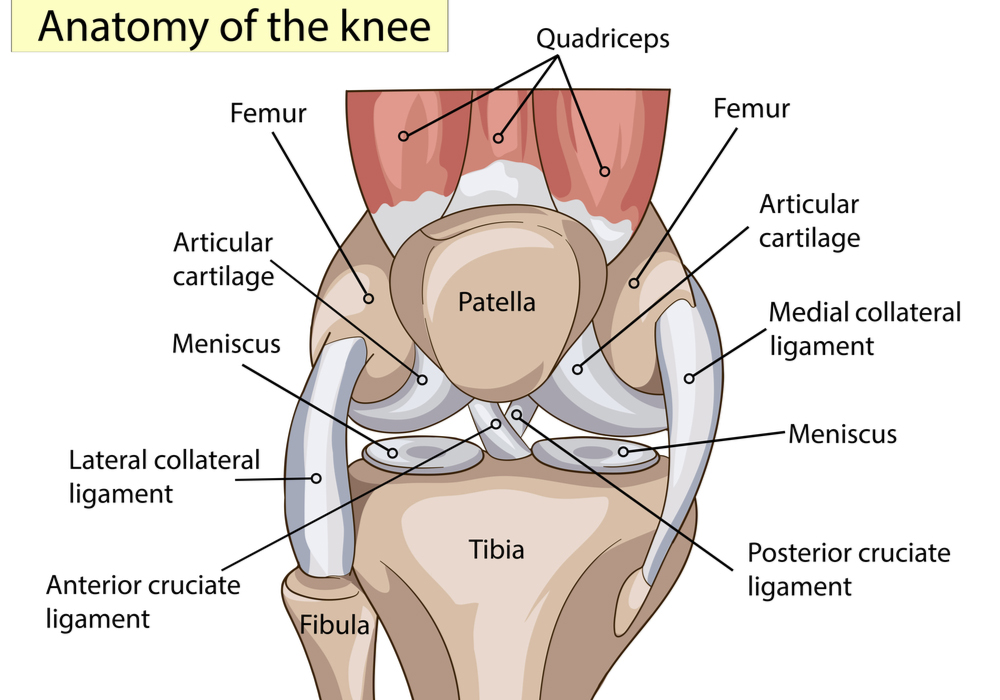
The knee joint is stable because four bones make up the knee anatomy. These bones include:
- Femur (thigh bone)
- Patella (kneecap)
- Tibia (shin bone)
- Fibula (small bone alongside shin bone)
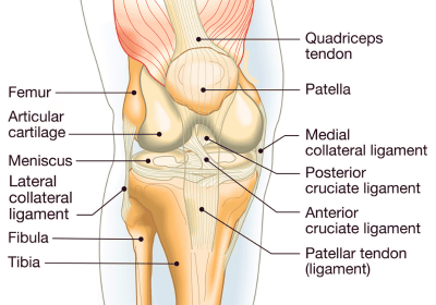
The femur forms the upper section of the knee, while the patella connects two tendons so we can bend the knee. The tibia and fibula make up the lower section of the knee. Together, these bones comprise three compartments within the knee: medial, lateral, and patellofemoral.
Knee Cartilage:
There are two types of cartilage in the knee joint that are equally important.
- Articular cartilage: This is what allows the knee to glide easily, free from friction. The slippery, shiny layer of tissue is found at the ends of each knee bone and is also attached to the back of the patella. This is the shiny white surface at the end of a chicken bone.
- Meniscus: This is the rubbery, shock absorber of the knee that keeps you balanced. The meniscus consists of two half-moon-shaped cartilage segments. The medial meniscus is found on the inside of the knee, while the lateral meniscus is located on the outside of the knee joint.
Knee Ligaments:
The knee anatomy consists of four major ligaments that are responsible for holding the knee bones together through activity and for keeping them stable. They are:
- Anterior Cruciate Ligament (ACL) – The ACL is the most commonly injured ligament within the knee joint, especially among athletes. Located in the middle of the knee, it connects the back of the thigh bone to the front of the shin bone, preventing the shin bone from moving forward. This ligament is important for sudden movements, like cutting and pivoting. Untreated ACL tears increase the risk of damaging surrounding structures, like the meniscus and articular cartilage.
- Posterior Cruciate Ligament (PCL) – The PCL is located within the knee joint behind the ACL and connects the back of the thigh bone to the front of the shin bone to form an ”X” within the knee. The PCL’s main job is to keep the tibia from moving backward in relation to the femur (thigh bone). PCL tears are more rare, due to the size and strength of this ligament. A forceful collision or other high impact accident, such as a major vehicle accident, can cause injury to the PCL.
- Medial Collateral Ligament (MCL) – This ligament is located on the inner side of the knee connecting the upper shin bone to the bottom of the thigh bone. Unlike the ACL and PCL, the MCL is technically outside the knee joint because it lives outside the protective capsule that lubricates the joint. If you play a sport with side-to-side motion, the MCL helps keep the knee stable. Pressure from outside the knee, forcing an extreme outward motion is usually the mechanism of injury to the MCL and is often seen in sports like tackle football.
- Lateral Collateral Ligament (LCL) Complex – Like the MCL, the LCL complex is technically outside the knee joint and also helps stabilize the knee during side-to-side movement. This ligament connects the top part of the fibula with the outside part of the lower thigh bone. The LCL is one of the key outer knee structures that control rotation and stability. A forceful collision or other high impact accident can cause an LCL injury.
Knee Tendons (Extensor Mechanism):
The tendons of the knee are similar to ligaments, but instead of connecting bones to bones, they connect muscles to bones.
- Patella tendon – This is the tendon that helps keep the patella in place by connecting the shin bone to the knee cap. It can be used as a graft site for ligament reconstruction. It can also tear in the setting of an injury and be repaired.
- Quadriceps tendon – It is one of the largest tendons within the knee anatomy and works with muscles and other tendons to help straighten the knee. It connects the front of the thigh to the kneecap. It can be used as a graft site for ligament reconstruction. It can also tear in the setting of an injury and be repaired.
- Pes Anserine – This is the area at the inner side of the knee near the top of the shin bone where three hamstring tendons come together. It can be used as a graft site for ligament reconstruction. It can also be a site of inflammation and irritation.
Muscles in the Knee:
The main structure of the knee anatomy doesn’t technically contain any muscles, but three nearby muscle groups do play a major role in providing knee strength, flexibility, and stability.
- Hamstring muscles: Four muscles found on the back of the thigh that are responsible for bending the knee.
- Quadriceps muscles: These four muscles help straighten the knee. They are located on the front of the thigh.
- Gluteal muscles: Known as the “glutes” or the “buttocks,” these four muscles include one of the largest and strongest muscles in the body, the gluteus maximus.


