Hip Specialist
The complex anatomy of the hip gives it an incredible range of motion, but can also leave it susceptible to injury. Selecting a specialist with experience treating injuries of the hip anatomy is crucial to a positive outcome. Doctor Prem Ramkumar, hip anatomy specialist, is located in Long Beach and serves patients in Los Angeles, Orange County, and surrounding Southern California areas. Contact Dr. Ramkumar’s office today!

What is the anatomy of the hip?
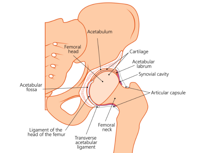
The hip joint connects the upper part of the leg (femur) to the torso (pelvis). The hip is surrounded by 20 muscles that attach to the knee, pelvis, and spine. As a result, any major issue originating from the hip can trigger back pain, knee pain, and even pain in the other hip. It is important to note an underlying spine issue can mimic hip findings or coexist with hip findings, more commonly known as Hip Spine Syndrome. Similarly, patients with back pain may have an underlying hip issue. The femur (thigh bone) and the acetabulum (pelvis bone) are the two bones that make up the hip anatomy. Often referred to as the ball and socket, the ball-shaped surface of the femoral head fits into a socket-like depression of the pelvic bone. Smooth cartilage connects the bones, stabilizing the joint and making it easy to move. Dr. Prem Ramkumar, hip anatomy specialist, is located in Long Beach and serves patients in Los Angeles, Orange County, and surrounding Southern California areas.

What makes up the hip joint?
Many parts of the hip anatomy are critical for daily activities like walking, running, and climbing stairs. If any part is injured, it can cause pain and affect the stability of the entire joint. Important hip anatomy structures include:
- Articular Cartilage – Allows the bones to move smoothly against each other.
- Synovial Membrane – Produces fluid that lubricates the cartilage and eliminates friction during movement.
- Bursa – Provides cushioning between muscles, tendons, and bones.
There are also key muscles in the hip anatomy responsible for helping the joint move smoothly and pain-free. They include:
- Gluteal or Abductor Muscles located on the side of the hip. (Include the Gluteus Minimus, Gluteus Medius, and Gluteus Maximus)
- Iliopsoas Muscle – Begins in the lower back and connects to the upper part of the femur in the front of the hip.
- Adductor Muscles – Muscles of the inner thigh that pull the leg inward toward the opposite leg and also help to flex and extend the hip.
- Quadriceps – Four muscles on the front of the thigh that run from the pelvis and hip joint down to the knee.
- Hamstrings – Muscles on the back of the thigh that run from the hip and pelvis to just below the knee.
Two major nerves and one artery also make up the hip anatomy:
The sciatic nerve is the longest and widest nerve in the human body. It runs down the back of the hip. The femoral nerve controls the muscles to straighten the leg and move the hips. It is at the front of the hip near the groin. The femoral artery is the largest artery in the thigh, supplying blood to the lower portion of the body.
Where exactly is hip pain?
Many do not realize that pain classically originating from the hip joint is typically in the front of the thigh near the groin. Less commonly, it presents on the side of the high. Even less commonly, pain can present in the buttock. Pain that travels from the top of the thigh beyond the knee towards the lower leg, ankle, and foot is typically pain originating from the back. Pain on the side of the thigh may be pain from within the hip joint, but it could also be from the abductors that stabilize the hip and pelvis. Seeing a specialist in the hip and knee is critical to evaluate the source of your pain to best dictate the plan.
What hip conditions cause pain?
- Arthritis of the hip
- Snapping or squeaking hip
- Femoroacetabular Impingement (FAI)
- Abductor Tendon Tears
- Psoas Impingement
- Trochanteric Bursitis
- Version abnormalities
What is the Layer Concept of the Hip?
Developed by Dr. Bryan Kelly from the Hospital for Special Surgery where Dr. Ramkumar trained, the Layer Concept is a systematic means of determining which structures in the non-arthritic hip are the source of the pathology, which are the pain generators, and how to best implement treatment. The four layers are comprehensively described below, but this framework helps simplify the complex nature of the hip. Recognizing and attempting to understand these osseous (Layer 1), inert (Layer 2), contractile (Layer 3), and neuromechanical (Layer 4) relationships and differences, particularly as they related to osseous over-coverage and under-coverage, underscores the value of the Layer Concept. The complexity of the hip joint, its associated anatomy, and its mimicry of pain sometimes in the back or the knee is why seeing a comprehensive hip specialist like Dr. Ramkumar can be important.
In the initial diagnostic process of evaluating a non-arthritic hip, it is helpful to categorize hips as normal, structurally overcovered, or structurally under-covered. Dr. Ramkumar uses the center edge angle, neck-shaft angle, femoral version (rotation), or acetabular version (direction) as metrics to assess this.
| Layer | Name: Structure | Purpose | Pathology |
|---|---|---|
| Osteochondral: Femur Osteochondral: Acetabulum Osteochondral: Innominate |
Joint congruence, Arthrokinetic movement | Developmental Dysplasia, Dynamic Cam Impingement, Rim Impingement, Trochanteric Impingement, Femoral Version, Acetabular Version, Femoral Inclination, Subspine Impingement, Acetabular Profunda/Protrusio |
| Inert: Capsule Inert: Labrum Inert: Capsuloligamentous Complex Inert: Ligamentum Teres |
Static Stability | Labral Tear, Capsular Instability, Ligamentum teres tear, Adhesive Capsulitis |
| Contractile: Musculature crossing hip Contractile: Lumbosacral muscles Contractile: Pelvic floor |
Dynamic Stability | Hip flexor strain; Psoas impingement; Rectus femoris impingement; Medial Enthesiopathy; Adductor tendinopathy; Rectus abdominus tendinopathy; Posterior Enthesiopathy; Proximal hamstring strain; Lateral Enthesiopathy; Peri-trochanteric space; Gluteus medius tear; Hemi-pelvic Pubalgia; Anterior Enthesiopathy |
| Neuromechanical: Thoroco-lumbar mechanics Neuromechanical: Lower extremity mechanics Neuromechanical: Neuro-vascular structures Neuromechanical: Regional mechanoreceptors |
Communication, timing and sequencing of the kinematic chain | Neural, Mechanical, Nerve Entrapment, Referred Spine Pathology, Scoliosis, Foot structure and mechanics, Pubic symphysis dysfunction, sacroiliaci joint dysfunction, Pain syndromes, Neuromuscular Dysfunction, Osteitis Pubis |


