SoCal Hip Arthroscopy Surgeon
Hip Arthroscopy is a minimally invasive procedure to treat early stage hip arthritis, hip instability, labral tears and other conditions. Dr. Prem Ramkumar, has extensive training and experience performing hip arthroscopy and serves patients in Los Angeles, Orange County, and surrounding Southern California (SoCal) areas. Contact Dr. Ramkumar’s team today!

What is hip arthroscopy?
Hip Arthroscopy is a highly specialized and minimally invasive procedure that allows Dr. Ramkumar to see inside the hip joint and reshape or reconstruct it with the help of a unique and small camera. This is reserved for patients with hip issues and little to no arthritis.
Hip arthroscopy is the gold standard operation for treating femoroacetabular impingement but is increasingly used in the treatment of patients with borderline hip dysplasia. How we approach, treat, and manage patients with non-arthritic hip conditions has changed dramatically over the last 10 years. Dr. Prem Ramkumar, hip arthroscopy surgeon, has extensive training and experience evaluating non-arthritic hip conditions and in performing this procedure. He is located in Long Beach and serves patients in Los Angeles, Orange County, and surrounding Southern California areas.
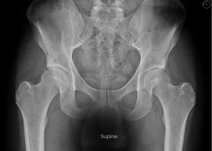
What is the hip joint?
The hip joint connects the upper part of the leg to the lower torso. It is surrounded by 20 muscles that attach to the knee, pelvis, and spine. As a result, any major issue originating from the hip joint can cause back pain, knee pain, and pain in the other hip. It is important to note an underlying spine issue can mimic hip findings or coexist with hip findings. The femur (thigh bone) and the pelvis (hip bone) are the two bones that make up the joint. Often referred to as the ball and socket, the ball-shaped surface of the femoral head fits into a cup-like depression of the pelvic bone. Smooth cartilage connects the bones, stabilizing the joint and making it easy to move.
Although the bony anatomy of the hip is a relatively simple structure to comprehend and visualize as a ball-and-socket joint, its surrounding structures are highly complex. The labrum, hip capsule, 17 accompanying muscles (attaching in different directions and tensions), and surrounding anatomy make recognizing hip conditions – especially in patients age 50 or younger – can be very challenging. Patients with pain that originates from the hip joint can complain of groin pain, outer thigh pain, back pain, low back pain, and even knee pain! Unlike many orthopaedic conditions, hip pain may present with more subtle symptoms like stiffness, achiness, muscle tightness, or a dull pain. Most commonly, patients will have pain getting in and out of a car or just sitting. There may be some catching and clicking as well.
What is femoroacetabular impingement syndrome (FAI syndrome)?
General Overview of FAI Syndrome
FAI syndrome is a constellation of symptoms originating from the repetitive contact between an abnormal bony growth on the femur (ball), acetabulum (socket), or both.
This bony overgrowth is caused by an injury during adolescence, a unique time in a person’s life when both the growth plate of the femur and the acetabulum (at the apophysis, where a key muscle of the quadriceps muscle originates) are capable of experiencing injury – and responding with abnormal bone creation. The abnormal bone deposited during this time period results in a decreased space with which the hip can move (stiffness, tightness, limited rotation) and injury to the immediately surrounding structures – such as the labrum and articular cartilage of the hip – resulting in pain. The constellation of symptoms resulting from this impingement between the femur and acetabulum within the hip give the name femoroacetabular impingement syndrome (FAI).
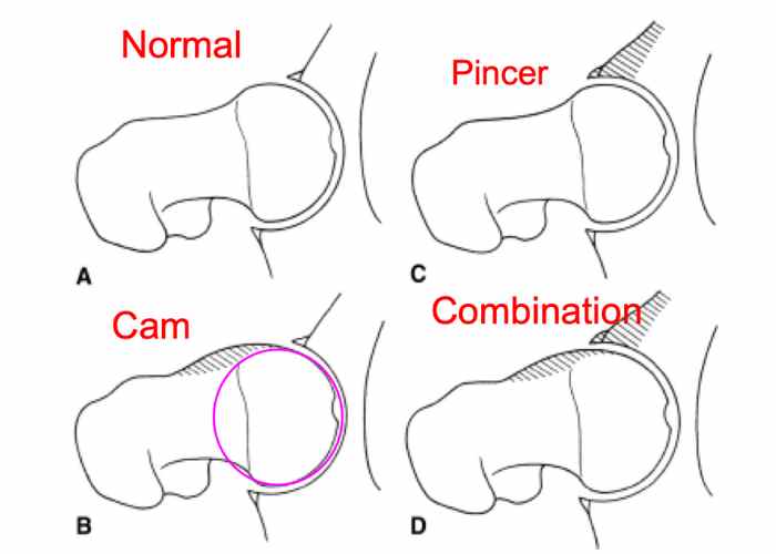
There are several distinct types of impingement. Patients may be diagnosed with one specific type, or a combination of these conditions:
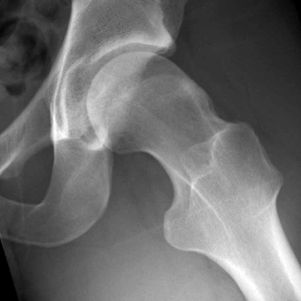
- Cam Impingement: This type of hip impingement occurs because of a misshaped femoral head and neck junction. If the ball of the femur is not round and smooth, it cannot rotate correctly inside the socket (acetabulum) – Cam deformities of the hip are often referred to as placing a square peg in a round hole. This anatomic abnormality causes excessive grinding against the cartilage inside the joint and can tear the labrum.
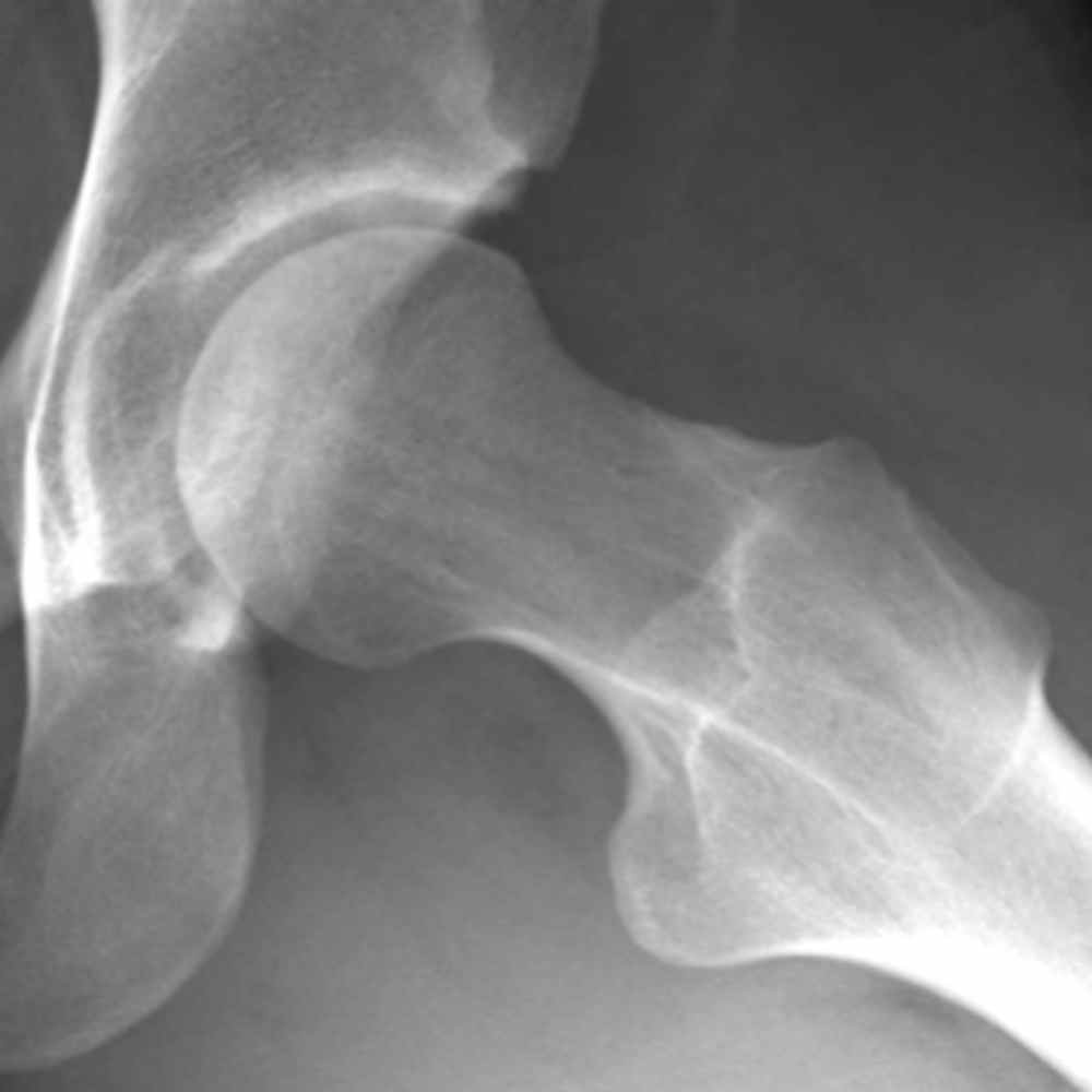
- Pincer Impingement: This type of hip impingement occurs when the front rim of the hip socket (called the acetabulum) sticks out too far. The neck of the femur (thigh bone) bumps into the protruding rim of the socket during normal hip flexion. This may cause damage to the articular cartilage and may crush the labrum.
- Cam and Pincer Impingement: Some patients exhibit a combined impingement where both the pincer and cam impingements are present.
What are the symptoms of femoroacetabular impingement?
Patients typically complain of:
- Sharp, ongoing pain in the groin area, especially when the hip is flexed and internally rotated (turned inward).
- Pain that radiates to the outside of the hip.
- Pain that radiates to the buttock
- Pain that can vary from a dull ache to stabbingly intense.
- A limp when starting to walk.
- Pain with sitting (classic finding)
- Getting in and out of a car
- Stiffness and decreased range of motion
Who is a candidate for hip arthroscopy?
Who is a candidate for hip arthroscopy?
Hip arthroscopy is primarily reserved for non-arthritic conditions, primarily FAIS. In certain cases it may be used to address patients with borderline dysplasia, as well as patients with both FAIS and dysplasia. It is generally performed on higher demand patients who have failed to respond to conservative treatments, such as rest and physical therapy. This condition is typically confirmed with history, exam, imaging, and an injection that ameliorates the pain. Dr. Ramkumar may recommend hip arthroscopy to relieve pain and restore joint function. Other conditions that may warrant hip arthroscopy include:
- PVNS – Synovitis
- Removal of loose bodies
- Trochanteric Bursitis
- Iliopsoas tendonitis
How is hip arthroscopy performed?
Hip arthroscopy is performed either as an inpatient or outpatient procedure. Dr. Ramkumar will make a small incision near your hip and gently insert a small camera, attached to a long tube known as an arthroscope. Once the camera is inside the hip joint, Dr. Ramkumar is able to see the joint on a monitor. He then can diagnose and treat a wide range of conditions using miniature instruments. Since only a tiny incision is made during hip arthroscopy, recovery times are much faster than with traditional surgeries.
What can I expect before and after hip arthroscopy surgery?
Dr. Ramkumar emphasizes the importance of establishing clear expectations and providing comprehensive pre- and post-surgery education. This proactive approach not only alleviates anxiety but also leads to an enhanced overall surgical outcome.
Learn more >> Preoperative Assessment and Postoperative Guidelines for Hip Arthroscopy Surgery
What is the recovery process after hip arthroscopy?
Hip arthroscopy is the only operation in orthopedics intended to increase the range of motion of a joint. As such, the hip and surrounding muscles will need time to adjust from a prolonged abnormal state to one that now has newfound range of motion. Physical therapists are crucial in this process.
Most patients are able to walk normally after hip arthroscopy in about four weeks. As with any procedure, the exact time it will take to heal varies from person to person and also depends on what repairs were made to the hip joint. Dr. Ramkumar will want you to use crutches for about a week or two after surgery. After two weeks, you should be able to start progressively increasing your weight-bearing. Your motion will need to be restricted in the first 4 weeks, and we use a brace to help you with this.
- We use a blood thinner to limit blood clots, and it is critical you take this medication in the first four weeks even if you feel fine
- Typically Aspirin 81mg twice a day for two weeks
- In rare occasions, we may use Apixaban/Eliquis depending on your history
Selecting an orthopedic hip surgeon with experience performing hip arthroscopy is crucial to a positive outcome. Dr. Prem Ramkumar is located in Long Beach and serves patients in Los Angeles, Orange County, and surrounding Southern California (SoCal) areas.
")
Hip Arthroscopy Testimonial by Stephanie K
Latest Hip Arthroscopy Research
 Predicting the Risk of Subsequent Hip Surgery Before Primary Hip Arthroscopy for Femoroacetabular Impingement Syndrome: A Machine Learning Analysis of Preoperative Risk Factors in Hip Preservation
Predicting the Risk of Subsequent Hip Surgery Before Primary Hip Arthroscopy for Femoroacetabular Impingement Syndrome: A Machine Learning Analysis of Preoperative Risk Factors in Hip Preservation